Computer-assisted surgical procedures with LASAK BioniQ implants

MUDr. Jiri Hrkal
- Dr. Jiri Hrkal earned his medical degree from the Faculty of Medicine at Charles University in Plzen, Czech Republic, establishing a strong foundation for his career in dentistry.
- Since 1992, Dr. Hrkal has operated his own private practice, focusing on dental implantology and implant prosthetics, developing extensive expertise in these specialized fields.
- Dr. Hrkal frequently lectures at training events both domestically and internationally. He shares his professional insights through academic reports and conducts lectures on advanced topics such as dynamic and static navigation, augmentation procedures, and the application of PRGF.
- He is a member of the IGZ and DGI, serves as vice-president of the Czech Society of Implantology, and holds a position on the board of the An-Institute DTMD University Luxembourg.
Anamnesis
A 76-year-old patient, in good health and a non-smoker, sought treatment at our practice for an unilaterally shortened dental arch in his right mandible. His primary request was for a fixed, implant-supported denture to restore functionality and aesthetics. After a comprehensive evaluation, including a CBCT scan and dental impressions, we developed a treatment plan tailored to the patient‘s needs. Our approach involved placing three BioniQ implants in the regions of teeth 45, 46, and 47. These implants would serve as the foundation for a fixed, screw-retained metal-ceramic dental bridge designed to provide a durable and natural- looking solution.
Clinical casebook
Planning of the implants and the surgical template, taking into account the available bone volume and suitable restoration: at 45 and 46, always a 10 mm long BioniQ Straight implant with Ø 3.5 mm and at 47 a 10 mm long, Conical BioniQ implant with Ø 4.0 mm.
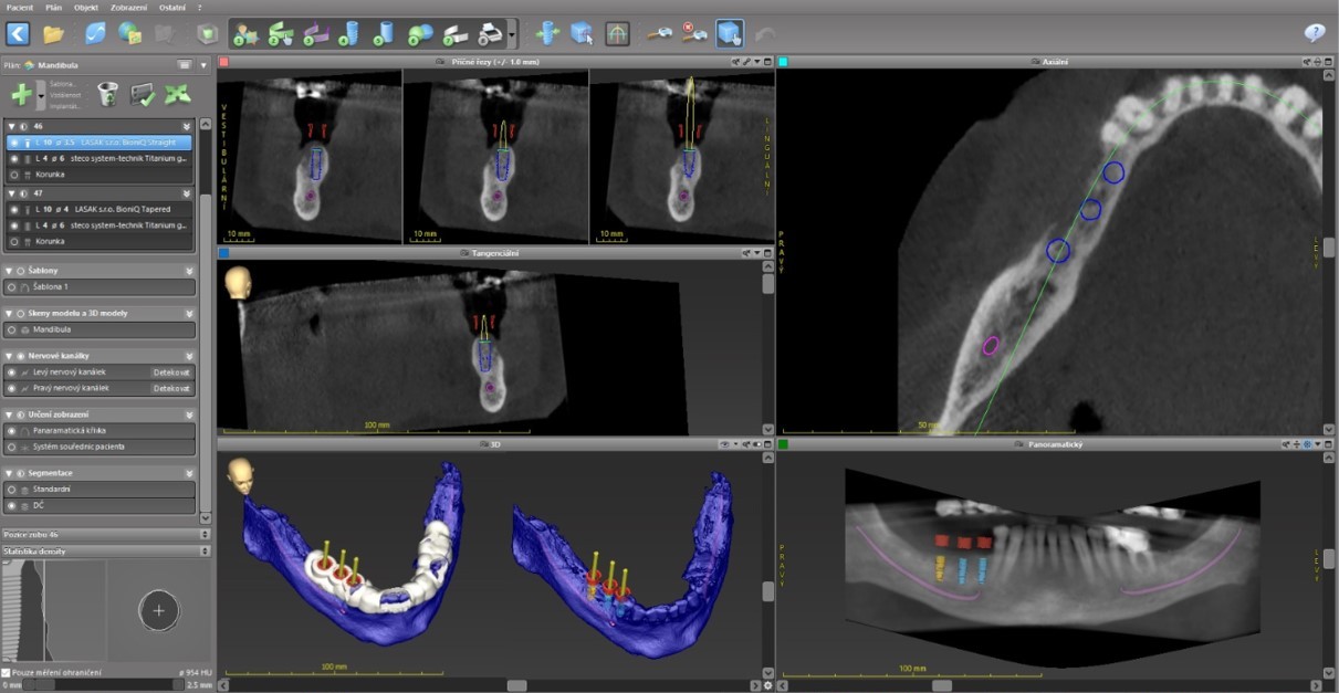
A 3D-printed tooth-supported surgical template was designed. The BioniQ instrument set for guided surgery was used for implant placement.

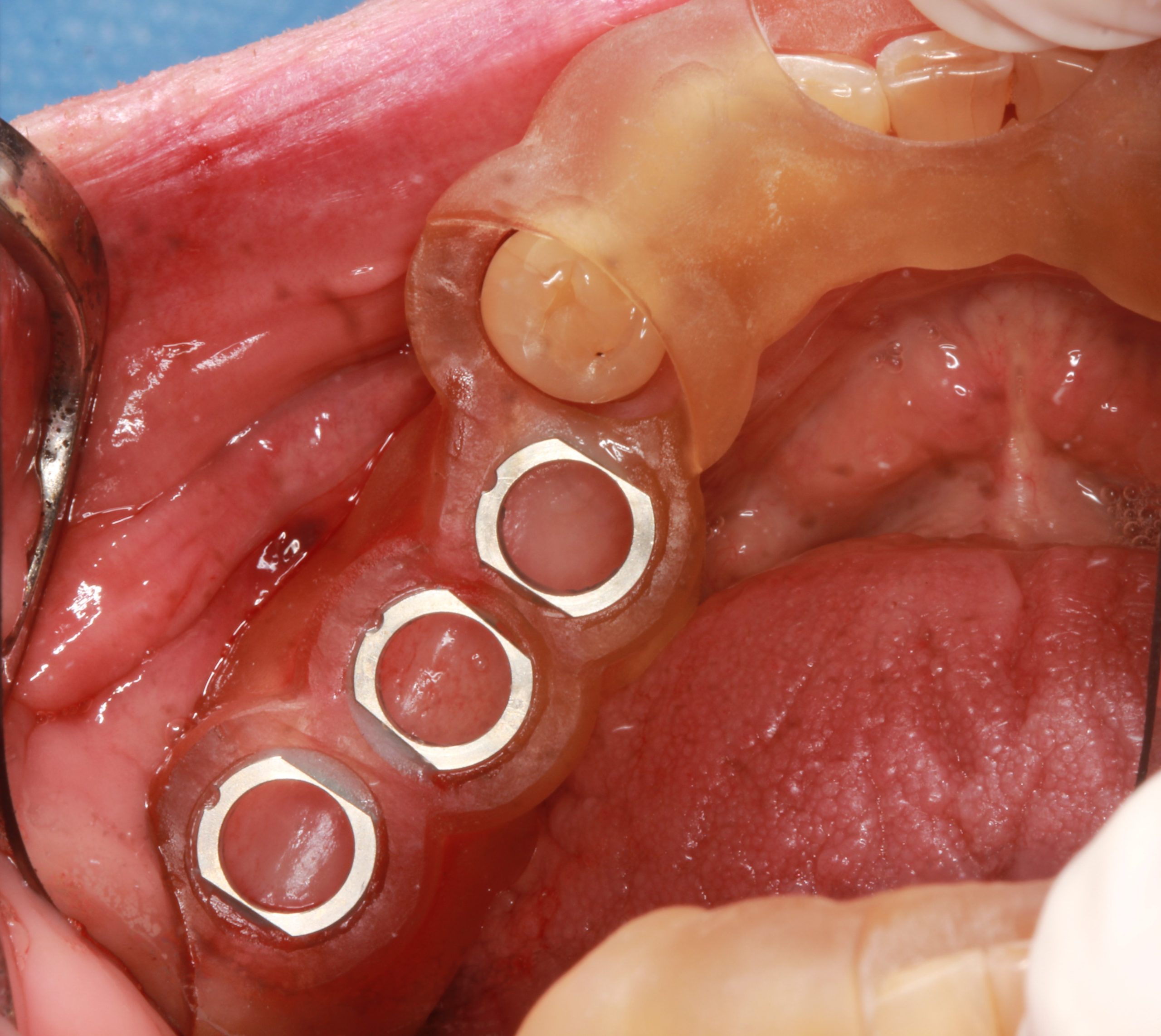
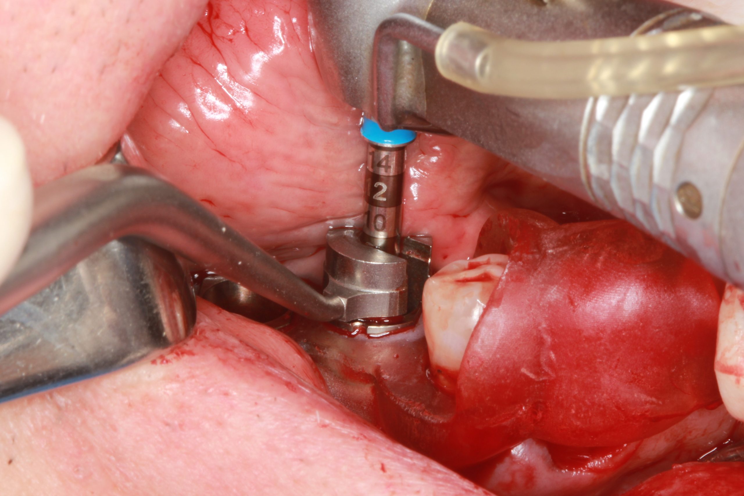
After rehearsing the exact fitting of the tooth-supported surgical template in the mouth, the implants were placed in the planned positions.
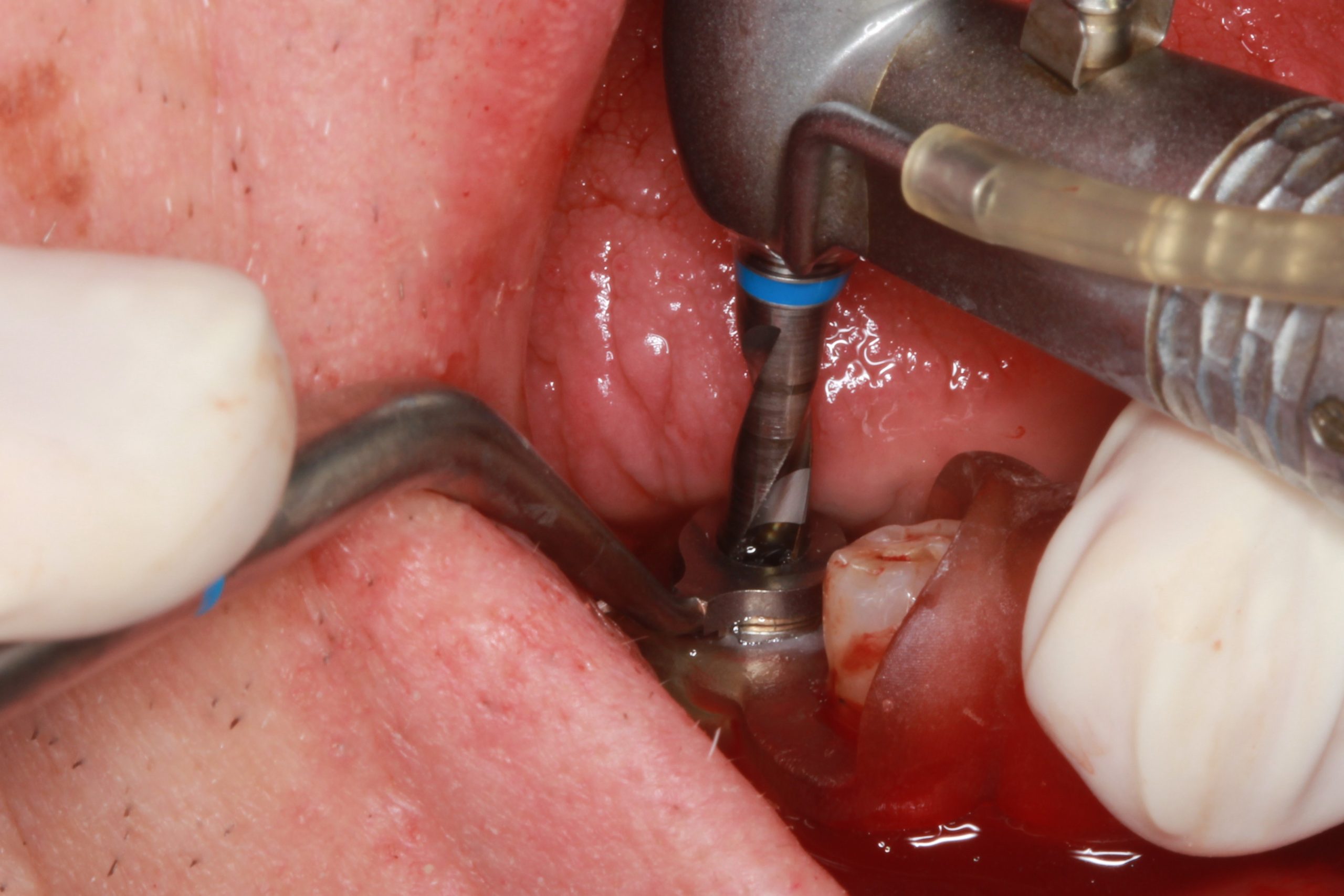
In accordance with the surgical protocol, the S2.9 guided drill with the required length and an S2.9 drill guide (with the same color strip) of the smallest diameter of 2.3 mm were used.
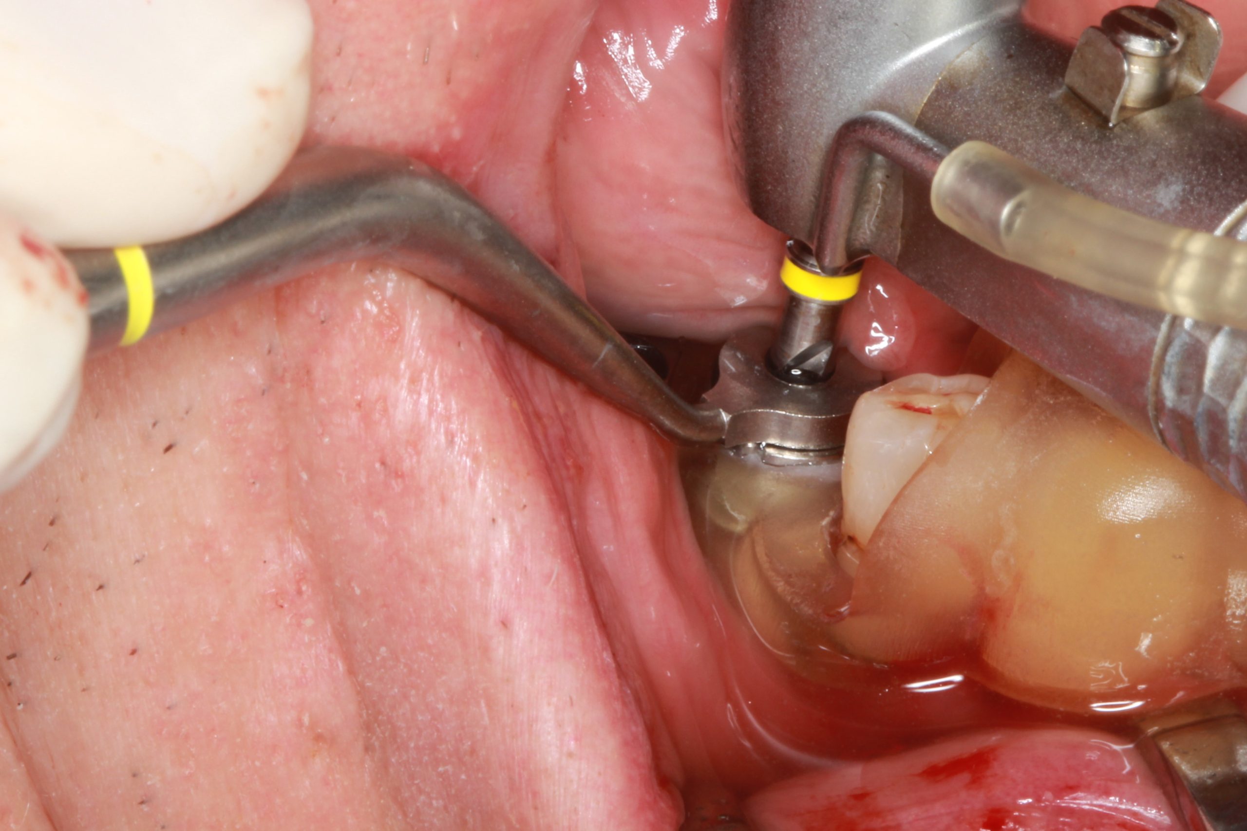
Then the treatment was continued with the drills and the drill guides for the appropriate implant diameter.
The preparation was completed using guided countersinks and threadformers with C-guides. After the surgical template was removed, the implant was placed.
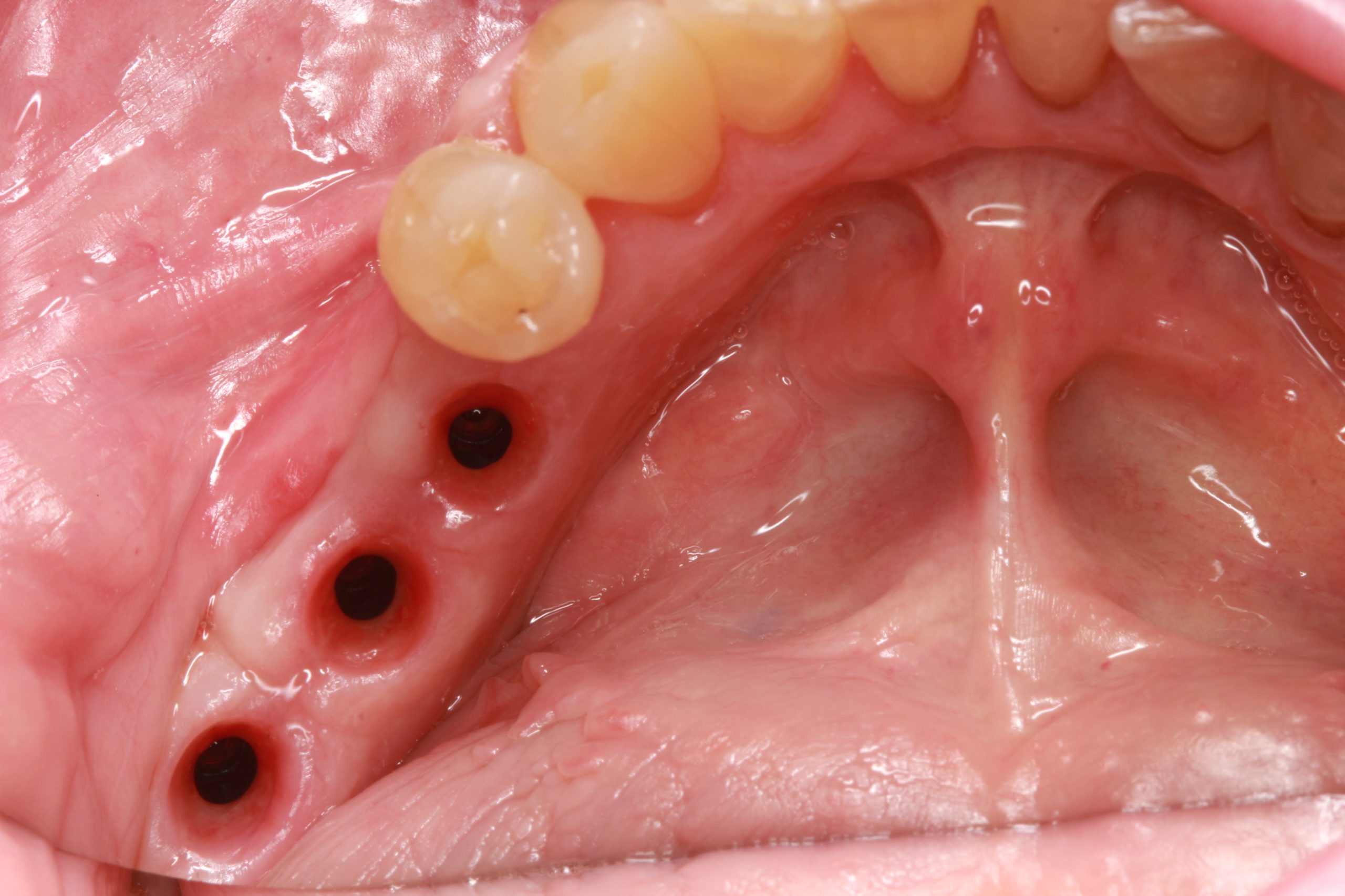
After three months, the implants were exposed, and the impression was taken.
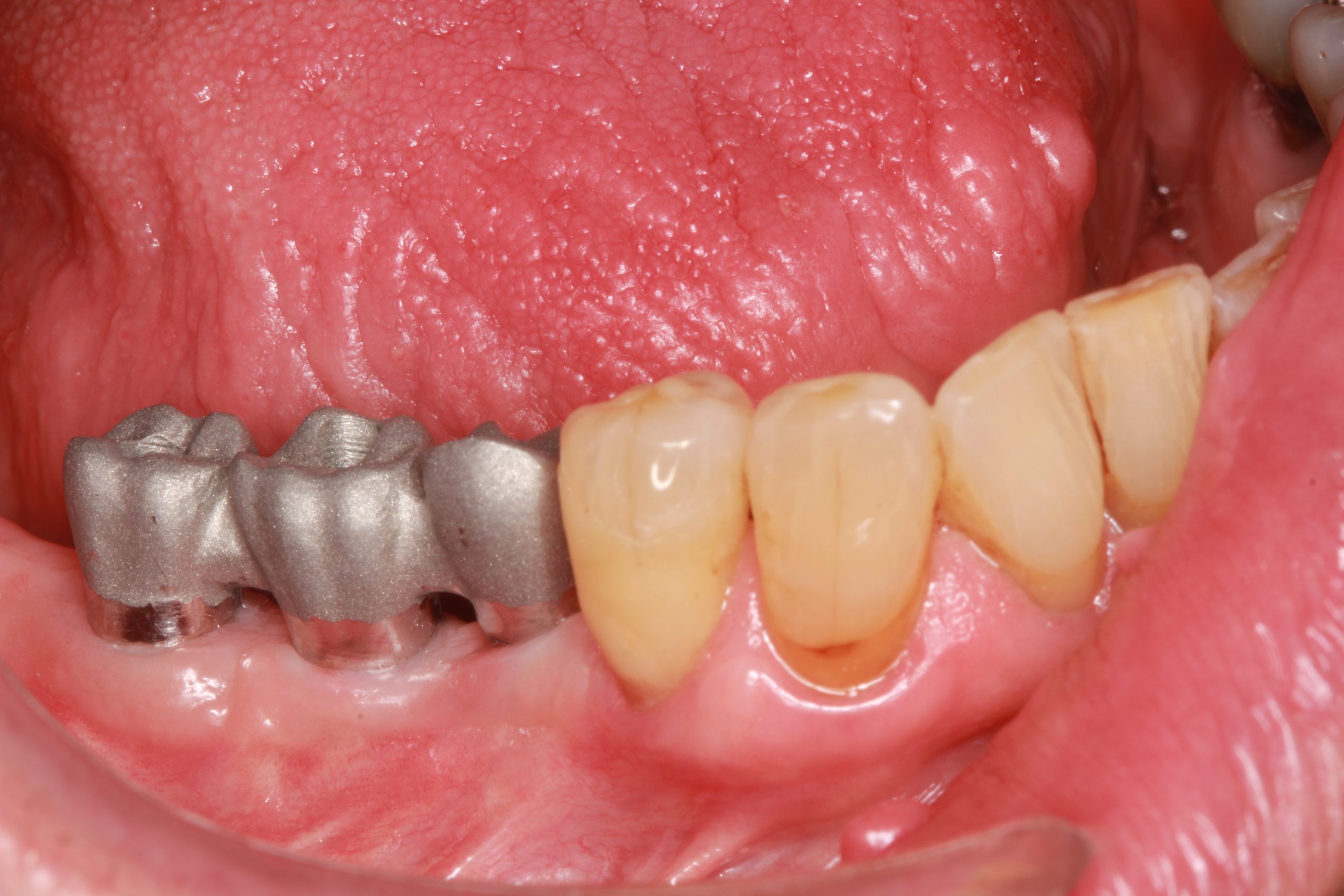
A fixed screw-retained metal-ceramic dental bridge was designed. The trial fitting of the superstructure shows that it fits correctly.
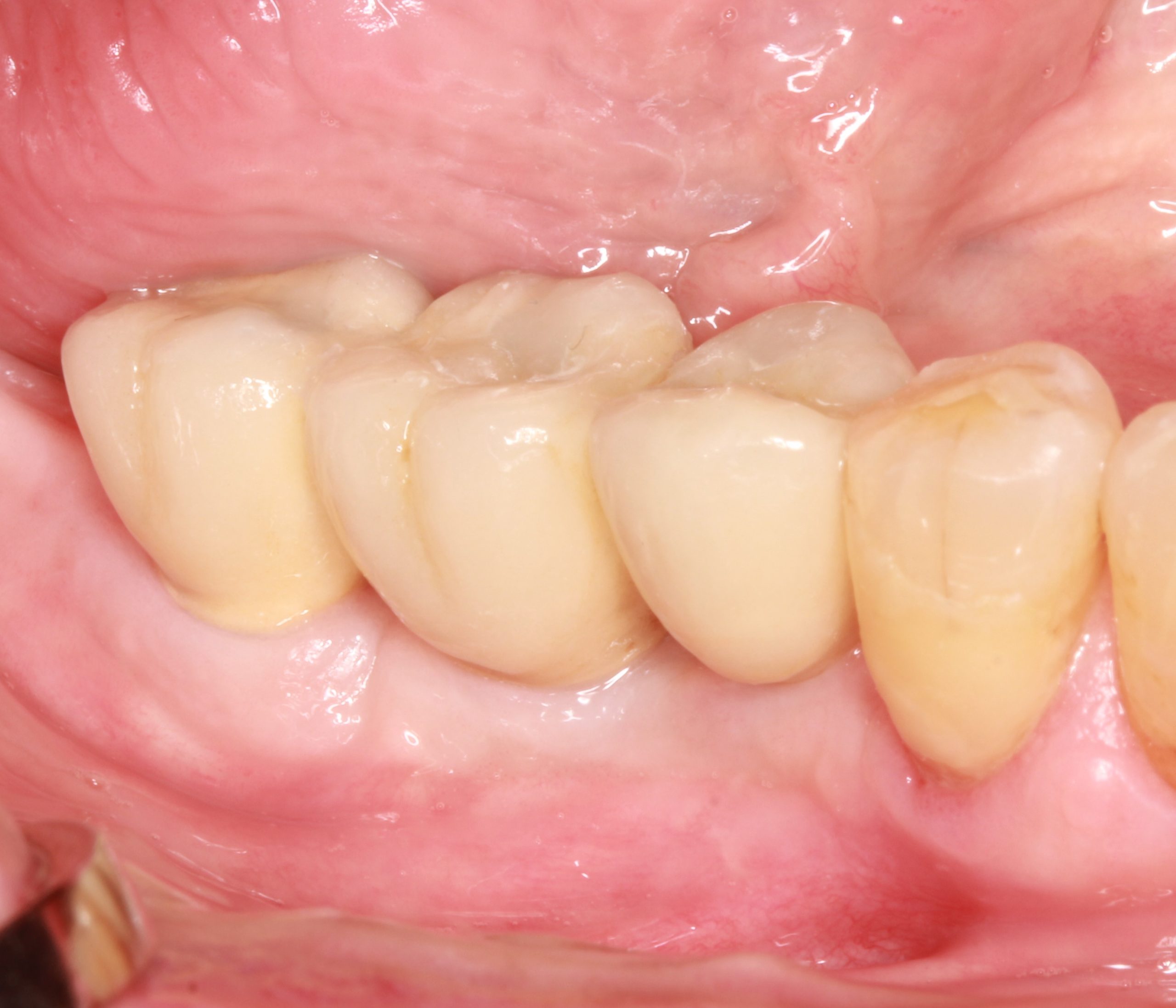
The final prosthesis was screwed into the patient‘s mouth. The patient’s expectations of a functional and esthetic result that is stable and sustainable in the long term were met.
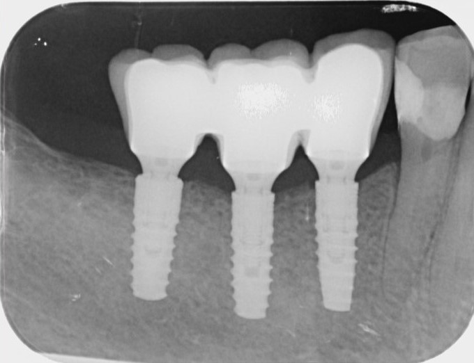
The X-ray check after fitting the final restoration.
Here you can download clinical casebook