Implantation with BioniQ pilot guided drilling in combination with dynamic navigation

MUDr. Jiri Hrkal
- Dr. Jiri Hrkal earned his medical degree from the Faculty of Medicine at Charles University in Plzen, Czech Republic, establishing a strong foundation for his career in dentistry.
- Since 1992, Dr. Hrkal has operated his own private practice, focusing on dental implantology and implant prosthetics, developing extensive expertise in these specialized fields.
- Dr. Hrkal frequently lectures at training events both domestically and internationally. He shares his professional insights through academic reports and conducts lectures on advanced topics such as dynamic and static navigation, augmentation procedures, and the application of PRGF.
- He is a member of the IGZ and DGI, serves as vice-president of the Czech Society of Implantology, and holds a position on the board of the An-Institute DTMD University Luxembourg.
Anamnesis
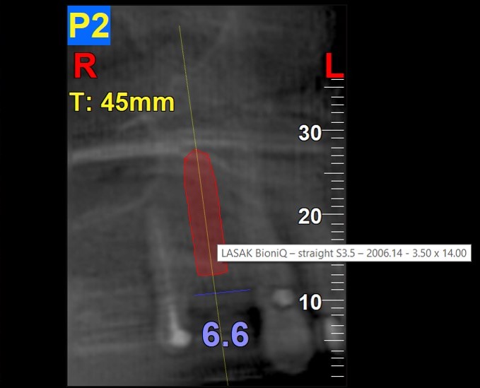
A 65-year-old female patient, in good health and a non-smoker, sought treatment at our practice. Her case required the removal of an unsatisfactory fixed restoration supported by teeth 17, 15, and 14, as well as the extraction of tooth 14. Teeth 17 and 15 had previously undergone endodontic treatment. After a thorough assessment, we developed a comprehensive treatment plan. This included placing a 14 mm long BioniQ Straight implant with a 3.5 mm diameter in the region of tooth 14. We also planned a fixed metal-ceramic bridge spanning teeth 17-15 and a metal-ceramic implant-supported bonded dental crown. Given the complexity of the case we decided on a combined approach using guided pilot drilling and dynamic navigation.
Clinical casebook
A CBCT scan of the maxilla was taken and then merged with the model scan using the coDiagnostiX software. A fixed metal-ceramic dental bridge on teeth 17-15 and a metal-ceramic implant-supported bonded dental crown for region 14 were planned and designed.
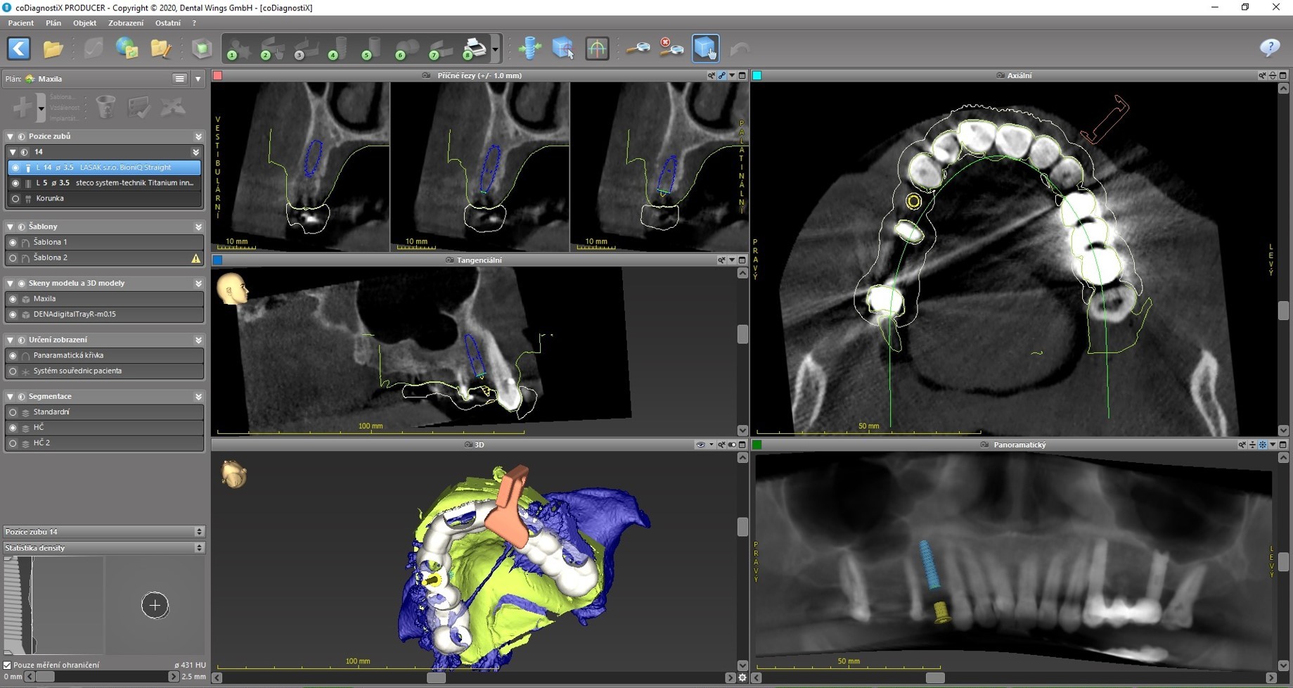
Based on the measurement of the bone volume, a 14 mm long BioniQ Straight implant with a diameter of 3.5 mm was planned for region 14.
In order to make the implant placement as precise as possible (given the limited bone supply and the implant length of 14 mm) and to avoid the nearby root of the neighboring tooth, a combined surgical template was designed.
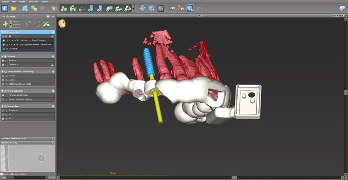
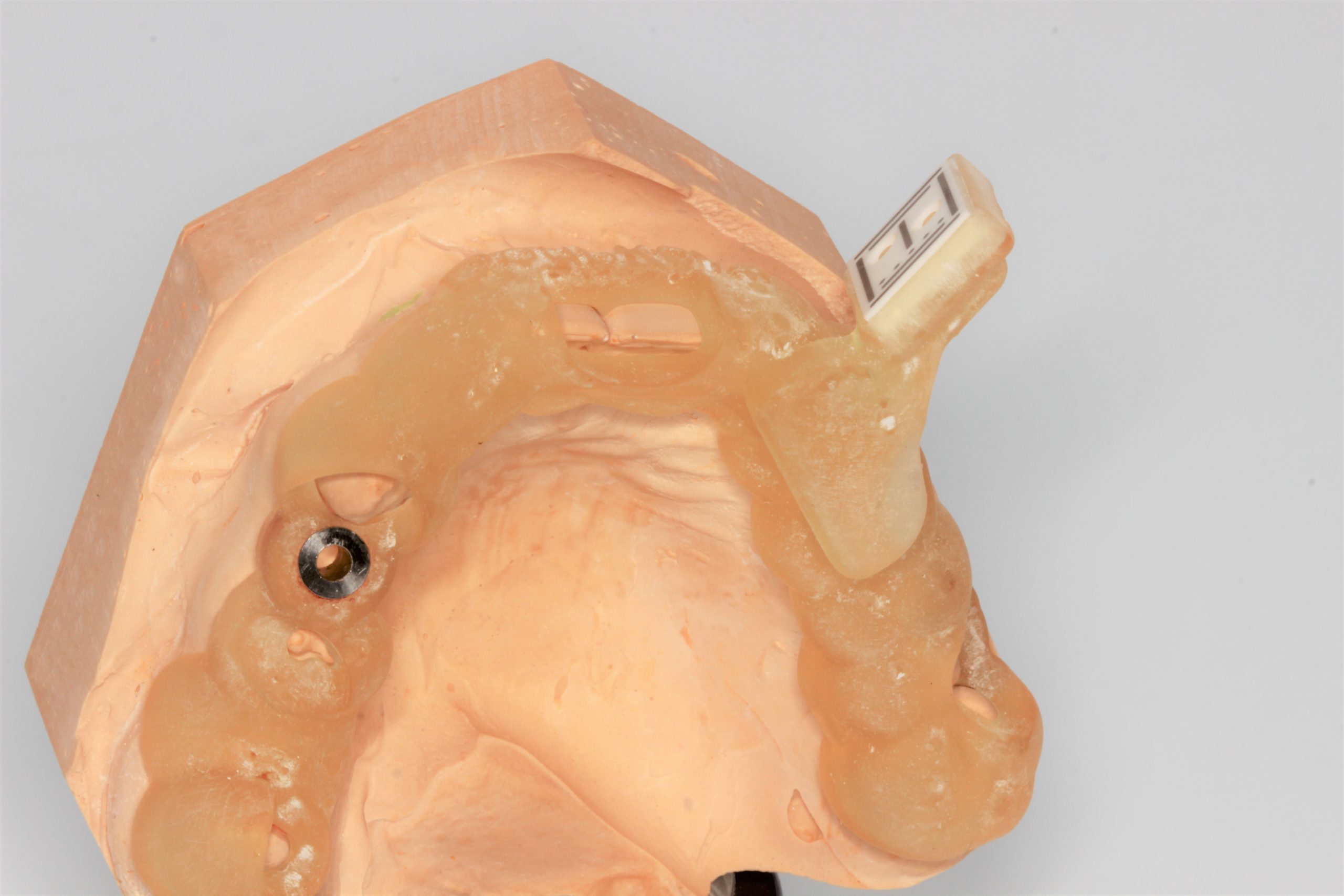
The tooth-supported surgical template contained a guide sleeve for the static template-guided pilot drilling and a holder for the dynamic navigation sensor. The template was produced using a 3D printer.
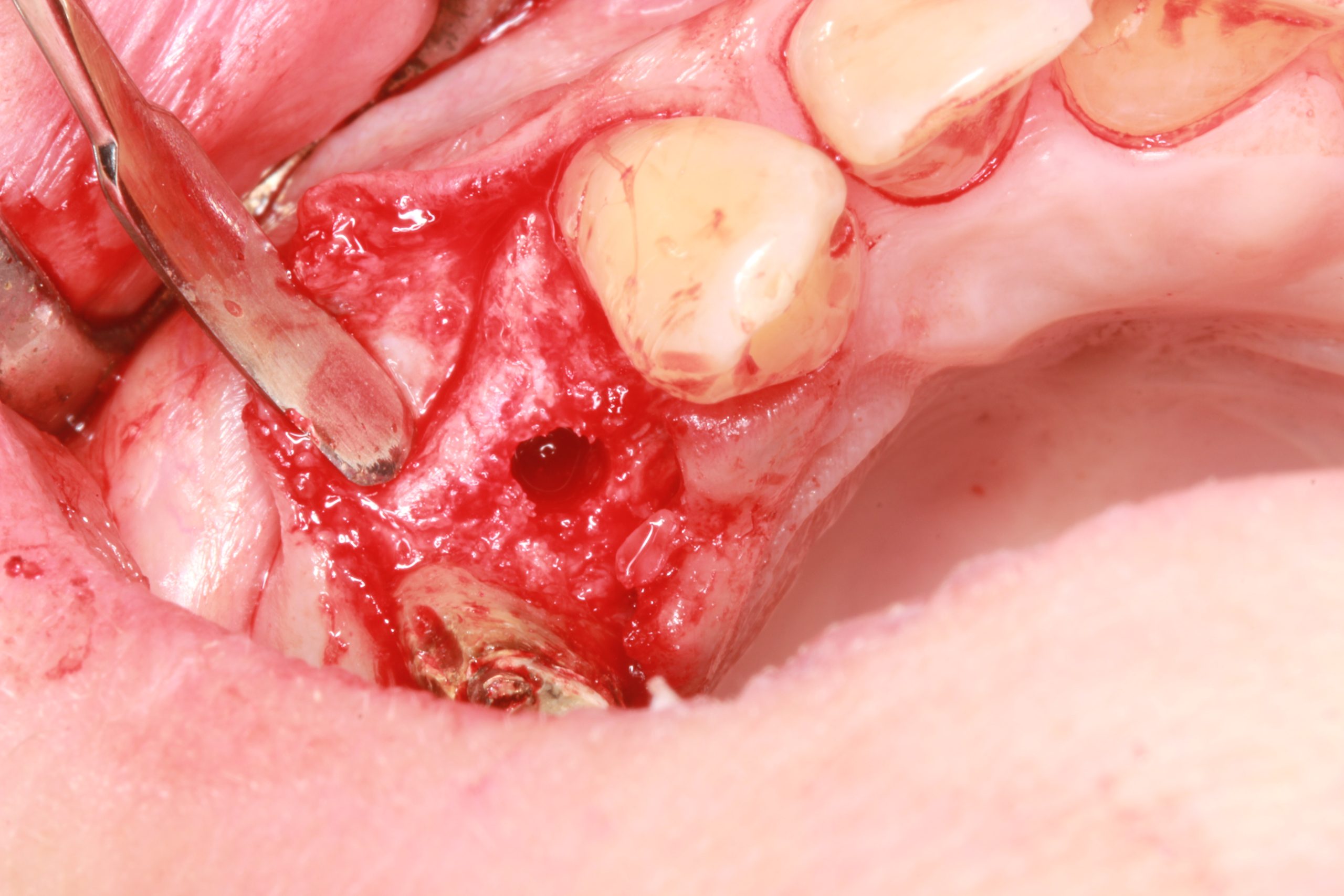
The implant bed preparation was started using the surgical template with the BioniQ S2.9 guided drill under the supervision of the DENACAM dynamic navigation system.
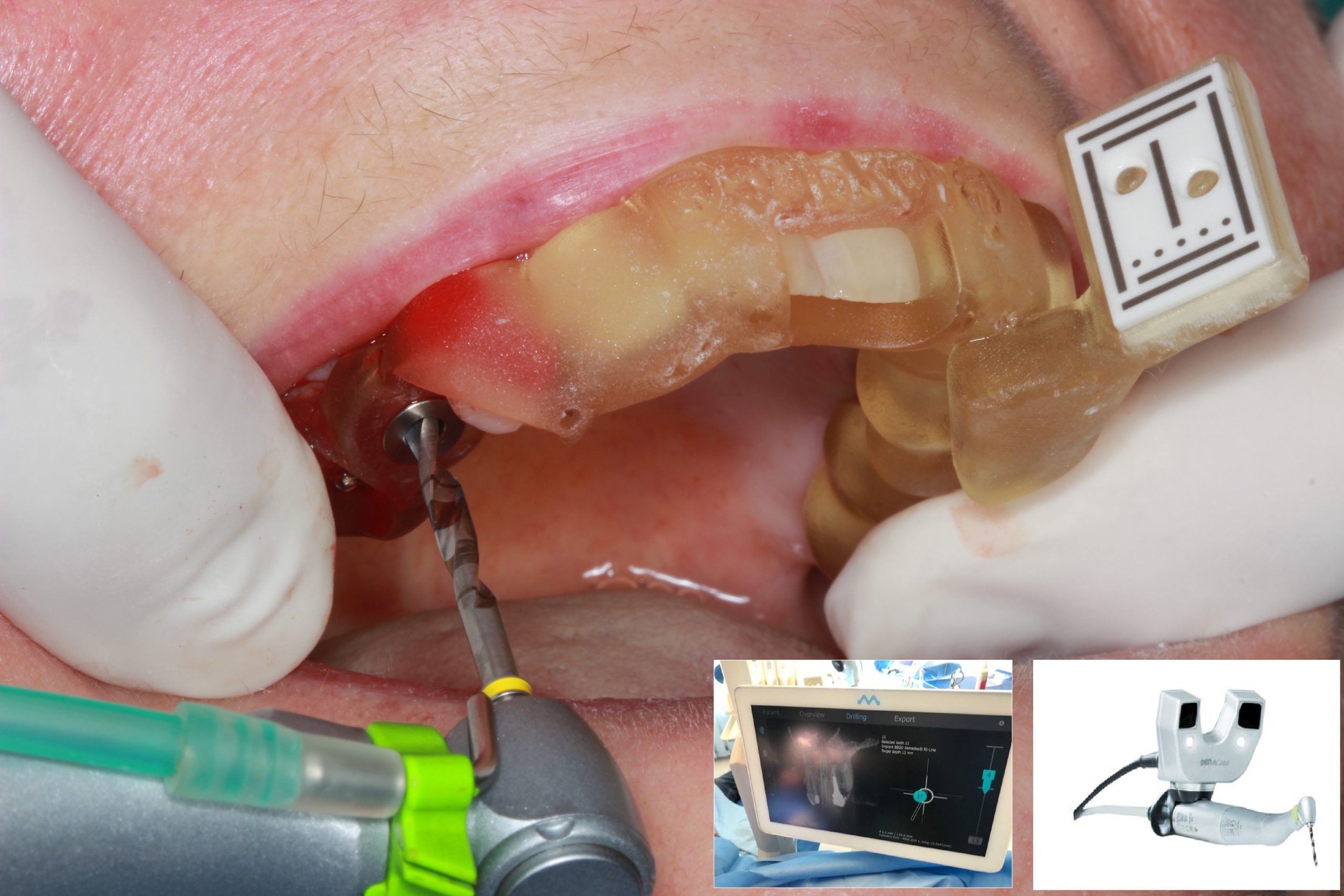
The template was removed and the preparation was completed with conventional instruments using the dynamic navigation system. The exact position of the planned implant was checked on the screen.
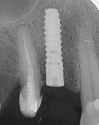
X-ray control image after the implantation. The implant was inserted as planned.
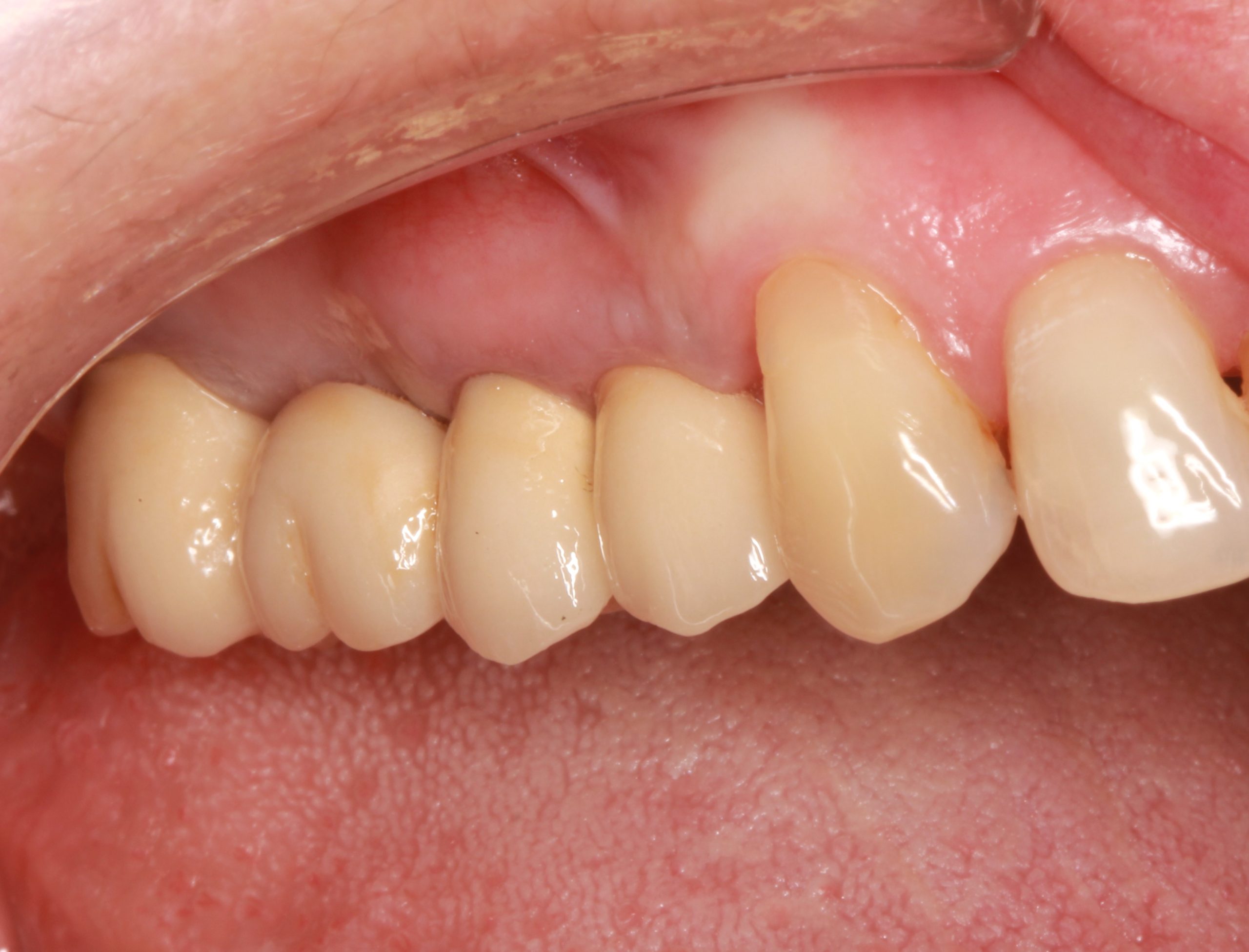
The final result after placing a fixed dental bridge on teeth 17–15 and an implant-supported dental crown on tooth 14.
Here you can download clinical casebook