BioniQ guided surgery in case of low bone volume

MUDr. Pavel Hyspler
- Dr. Pavel Hyspler completed his dental education at Charles University in Hradec Kralove, Czech Republic, laying the foundation for his distinguished career.
- He gained diverse experience during his residency, dividing his time between a private practice and the Department of Stomatology at Prague University Hospital.
- In 2009, Dr. Hyspler‘s expertise and leadership skills were recognized when he was appointed head of the Department of Stomatology at Military University Hospital Prague.
- Beyond his clinical responsibilities, Dr. Hyspler actively contributes to the field of dentistry through product development and research projects. He regularly shares his findings through professional reports and as a speaker at conferences both in the Czech Republic and internationally.
Anamnesis
A 52-year-old female patient sought treatment for complications with an implant at the site of tooth 36. The implant, placed approximately six years earlier at another practice, had begun to show signs of loosening. An OPG revealed significant bone loss around the implant, indicative of peri-implantitis. Given the extent of the damage, we decided to remove the failing implant and excochleate the defect. After a three-month healing period, we performed a CBCT scan to plan a new implant-supported restoration. After careful consideration of all available options, we determined that a tilted implant would be the most suitable approach. This implant was strategically inserted to bypass the mandibular canal, ensuring optimal positioning while preserving vital structures.
Clinical casebook
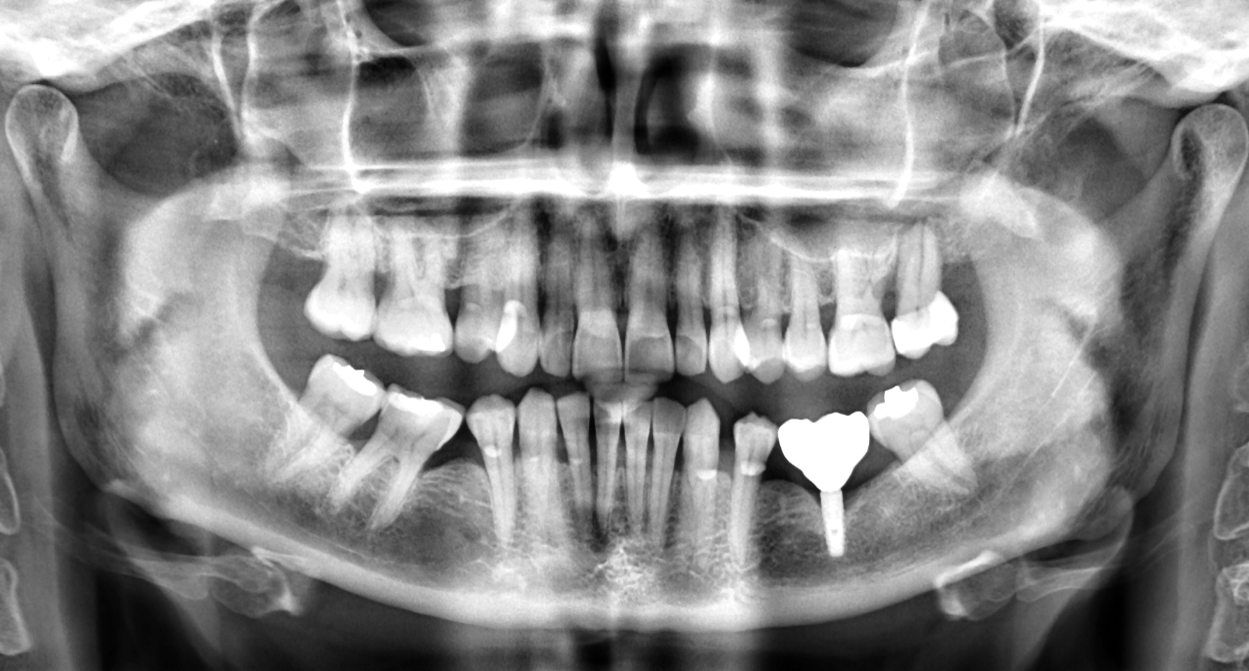
Initial radiograph before the start of treatment. The bone loss caused by peri-implantitis is visible.
CBCT scan three months after implant removal.

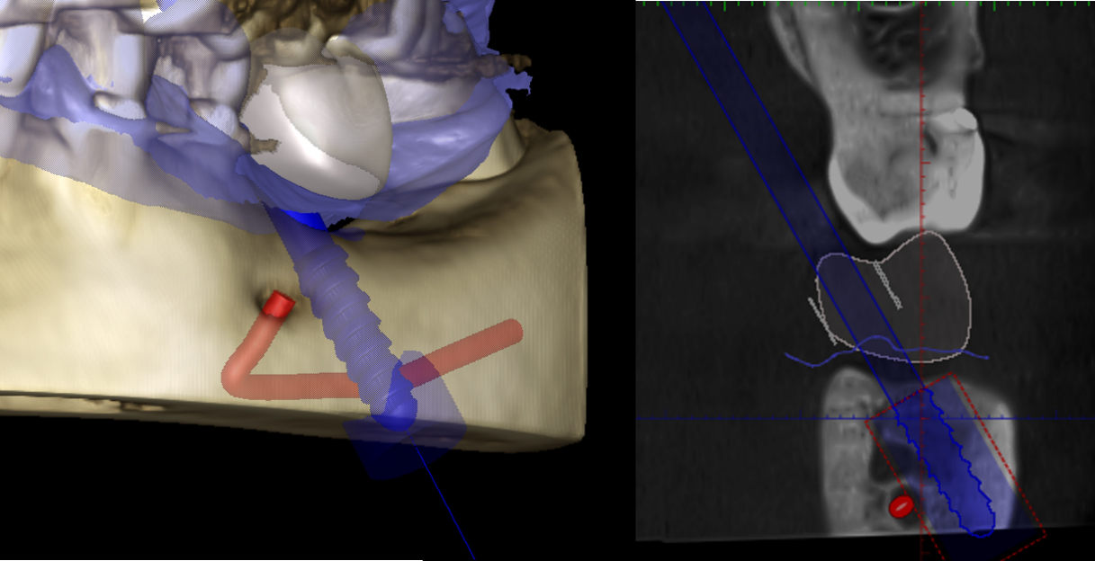
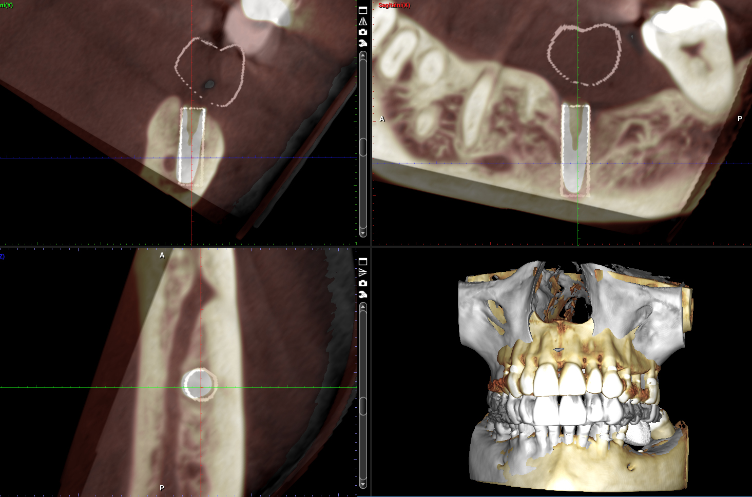
Planned position of the BioniQ implant near the mandibular canal.
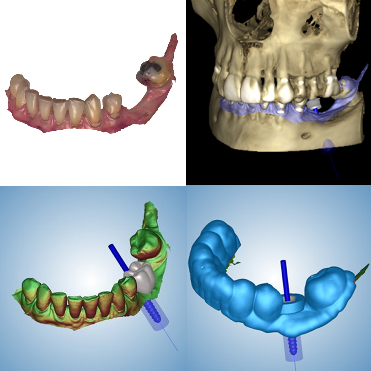
Design of a surgical template in the Romexis program, taking into account the requirements for implant position as well as a suitable restoration.
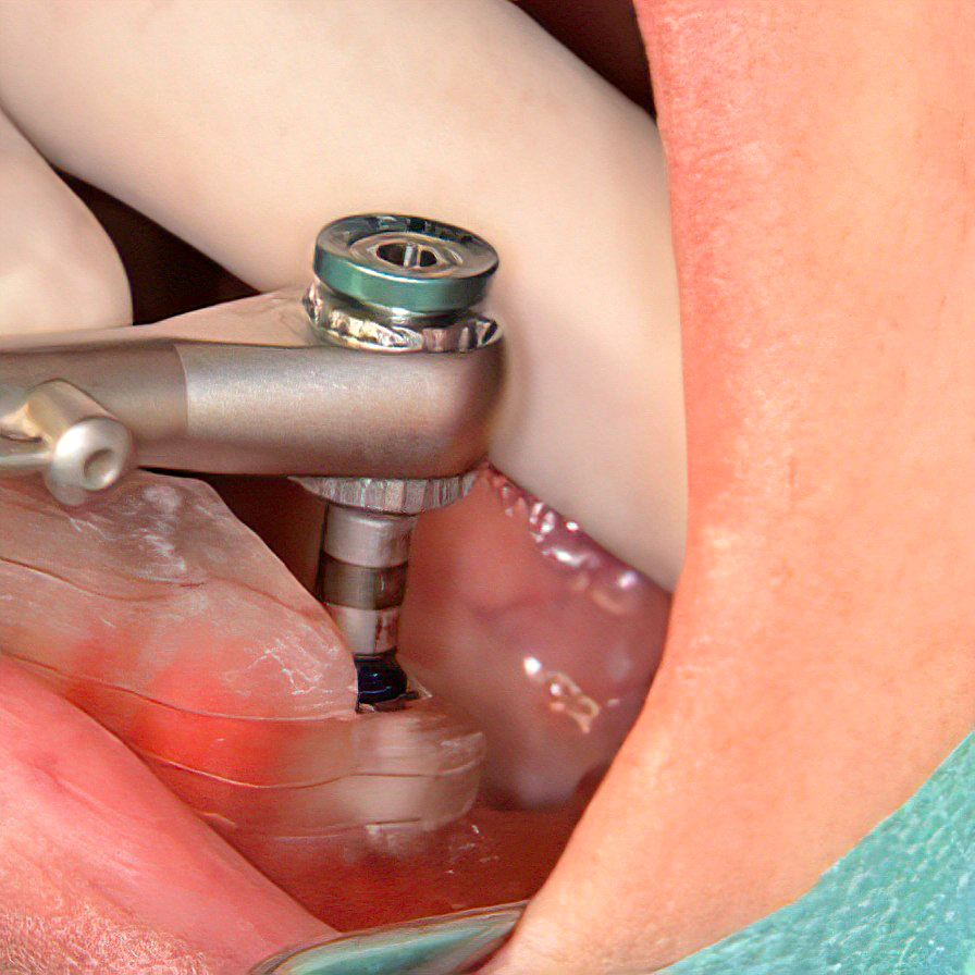
The BioniQ implant with a diameter of 3.5 mm and a length of 12 mm was inserted through a surgical template using an insertion wrench with marked offsets.
Accuracy of template guided implantation – the planned position is represented by a white cylinder. The implant was inserted according to plan with a clinically insignificant deviation.
The emergence profile of the mandible, scan of the mandible with scanbody, intraoral scan of the maxilla, and occlusion scan.

Design of the individual abutment and the dental crown in the exocad program. An individual abutment with an angled screw channel was planned for better access to the restoration.

Final dental prosthesis. The custom abutment was bonded into the zirconia crown in the laboratory.
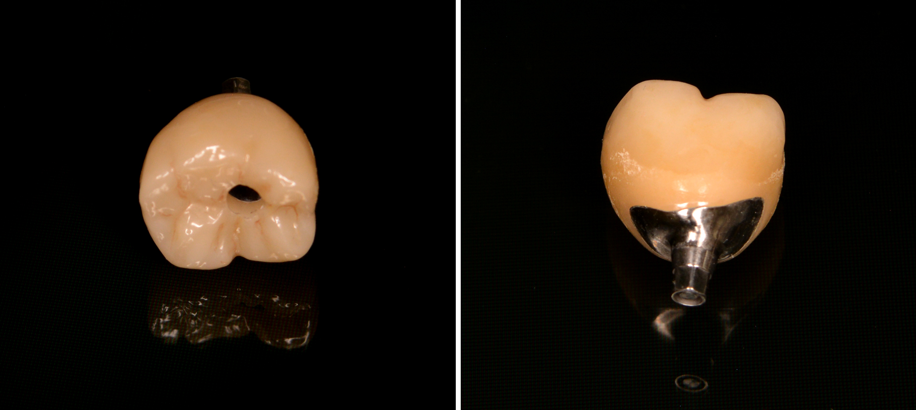
The final result after placing the dental crown at the site of tooth 36.

Here you can download clinical casebook